How is Thyroid Cancer Diagnosed?
Tests for Thyroid Cancer
Thyroid cancer may be diagnosed after a person goes to a doctor because of symptoms, or it might be found during a routine physical exam. If there is a reason to suspect you might have thyroid cancer, your doctor will use one or more tests to further research your suspicions. Signs and symptoms might suggest you have thyroid cancer, but you will need tests to confirm the diagnosis.
Medical History and Physical Exam
If you have any signs or symptoms that suggest you might have thyroid cancer, your health care professional will want to know your complete medical history. You will be asked questions about your possible risk factors, symptoms, and any other health problems or concerns. If someone in your family has had thyroid cancer, particularly medullary thyroid cancer or tumors called pheochromocytomas, it is important to tell your doctor, as you might be at high risk for this disease.
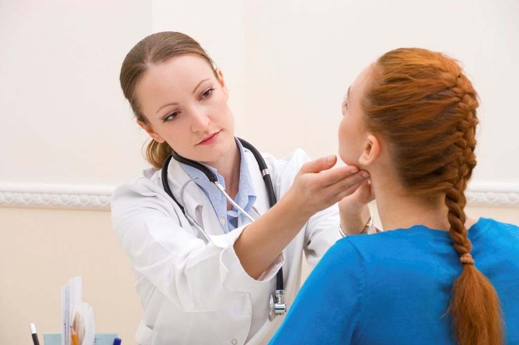
Your doctor will examine you to get more information about possible signs of thyroid cancer and other health problems. During the exam, the doctor will pay special attention to the size and firmness of your thyroid and any enlarged lymph nodes in your neck.
Biopsy
The actual diagnosis of thyroid cancer is made with a biopsy, in which cells from the suspicious area are removed and looked at under a microscope. However, this might not be the first test done if you have a suspicious lump in your neck. The doctor might order other tests first, such as blood tests, an ultrasound exam, or a radioiodine scan to get a better sense of whether you might have thyroid cancer. These tests are described below.
If your doctor thinks a biopsy is needed, the simplest way to find out if a thyroid lump or nodule is cancerous is with a fine needle aspiration (FNA) of the thyroid nodule. This type of biopsy can usually be done in your doctor’s office or clinic.
This test is generally done on all thyroid nodules that are big enough to be felt. This means that they are larger than about 1 centimeter across. Doctors often use ultrasound to see the thyroid during the biopsy, which helps make sure they are getting samples from the right areas. This is especially helpful for smaller nodules. FNA biopsies can also be used to get samples of swollen lymph nodes in the neck to see if they contain cancer.
Sometimes an FNA biopsy will need to be repeated because the samples didn’t contain enough cells. Most FNA biopsies will show that the thyroid nodule is benign. Rarely, the biopsy may come back as benign even though cancer is present. Cancer is clearly diagnosed in only about 1 of every 20 FNA biopsies.
Sometimes the test results first come back as “suspicious” or “of undetermined significance” if FNA findings don’t show for sure if the nodule is either benign or malignant. If this happens, the doctor may order tests on the sample to see if the BRAF or RET/PTC genes are mutated. Finding these changes makes thyroid cancer much more likely, and may also play a role in determining the best treatment for cancer.
Imaging Tests
Imaging tests may be done for a number of reasons, including helping find suspicious areas that might be cancer, to learn how far cancer may have spread, and to help determine if treatment is working. People who have or may have thyroid cancer will get one or more of these tests.
Ultrasound
Ultrasound uses sound waves to create images of parts of your body. For this test, a small, wand-like instrument called a transducer is placed on the skin in front of your thyroid gland. It gives off sound waves and picks up the echoes as they bounce off the thyroid. The echoes are converted by a computer into a black and white image on a computer screen. You are not exposed to radiation during this test.
This test can help determine if a thyroid nodule is solid or filled with fluid. It can also be used to check the number and size of thyroid nodules. How a nodule looks on ultrasound can sometimes suggest if it is likely to be cancer, but ultrasound can’t tell for sure.
For thyroid nodules that are too small to feel, this test can be used to guide a biopsy needle into the nodule to obtain a sample. Even when a nodule is large enough to feel, most doctors prefer to use ultrasound to guide the needle.
Ultrasound can also help determine if any nearby lymph nodes are enlarged because the thyroid cancer has spread. Many thyroid specialists recommend ultrasound for all patients with thyroid nodules large enough to be felt.
Radioiodine Scan
Radioiodine scans can be used to help determine if someone with a lump in the neck might have thyroid cancer. They are also often used in people who have already been diagnosed with differentiated thyroid cancer to help show if it has spread. Because medullary thyroid cancer cells do not absorb iodine, radioiodine scans are not used for this cancer.
For this test, a small amount of radioactive iodine (called I-131) is swallowed or injected into a vein. Over time, the iodine is absorbed by the thyroid gland. A special camera is used several hours later to see where the radioactivity is.
For a thyroid scan, the camera is placed in front of your neck to measure the amount of radiation in the gland. Abnormal areas of the thyroid that have less radioactivity than the surrounding tissue are called cold nodules, and areas that take up more radiation are called hot nodules. Hot nodules usually are not cancerous, but cold nodules can be benign or cancerous. Because both benign and cancerous nodules can appear cold, this test by itself can’t diagnose thyroid cancer.
After surgery for thyroid cancer, whole-body radioiodine scans are used to look for possible spread throughout the body. These scans become more sensitive if the entire thyroid gland has been removed by surgery because more of the radioactive iodine is picked up by any remaining thyroid cancer cells.
Radioiodine scans work best if patients have high blood levels of thyroid-stimulating hormone. For people whose thyroid has been removed, TSH levels can be increased by stopping thyroid hormone pills for a few weeks before the test. This leads to low thyroid hormone levels (hypothyroidism) and causes the pituitary gland to release more TSH, which in turn stimulates any thyroid cancer cells to take up the radioactive iodine. A downside of this is that it can cause the symptoms of hypothyroidism, including tiredness, depression, weight gain, sleepiness, constipation, muscle aches, and reduced concentration. One way to raise TSH levels without withholding thyroid hormone is to give an injectable form of thyrotropin before the scan. Because any iodine already in the body can affect this test, people are usually told not to ingest foods or medicines that contain iodine in the days before the scan. Radioactive iodine can also be used to treat differentiated thyroid cancer, but it is given in much higher doses. This type of treatment is described in the section Radioactive iodine (radioiodine) therapy.
Chest x-ray
If you have been diagnosed with thyroid cancer (especially follicular thyroid cancer), a plain x-ray of your chest may be done to see if the cancer has spread to your lungs.
Magnetic Resonance Imaging (MRI) Scan
Like CT scans, MRI scans can be used to look for cancer in the thyroid, or cancer that has spread to nearby or distant parts of the body. But ultrasound is usually the first choice for looking at the thyroid. MRI can provide very detailed images of soft tissues such as the thyroid gland. MRI scans are also very helpful in looking at the brain and spinal cord.
Blood Tests
Blood tests are not used to find thyroid cancer. But they can help show if your thyroid is working normally, which may help the doctor decide what other tests may be needed. They can also be used to monitor certain cancers.
Vocal Cord Exam (Laryngoscopy)
Thyroid tumors can sometimes affect the vocal cords. If you are going to have surgery to treat thyroid cancer, a procedure called a laryngoscopy will probably be done first to see if the vocal cords are moving normally. For this exam, the doctor looks down the throat at the larynx (voice box) with special mirrors or with a laryngoscope, a thin tube with a light and a lens on the end for viewing.